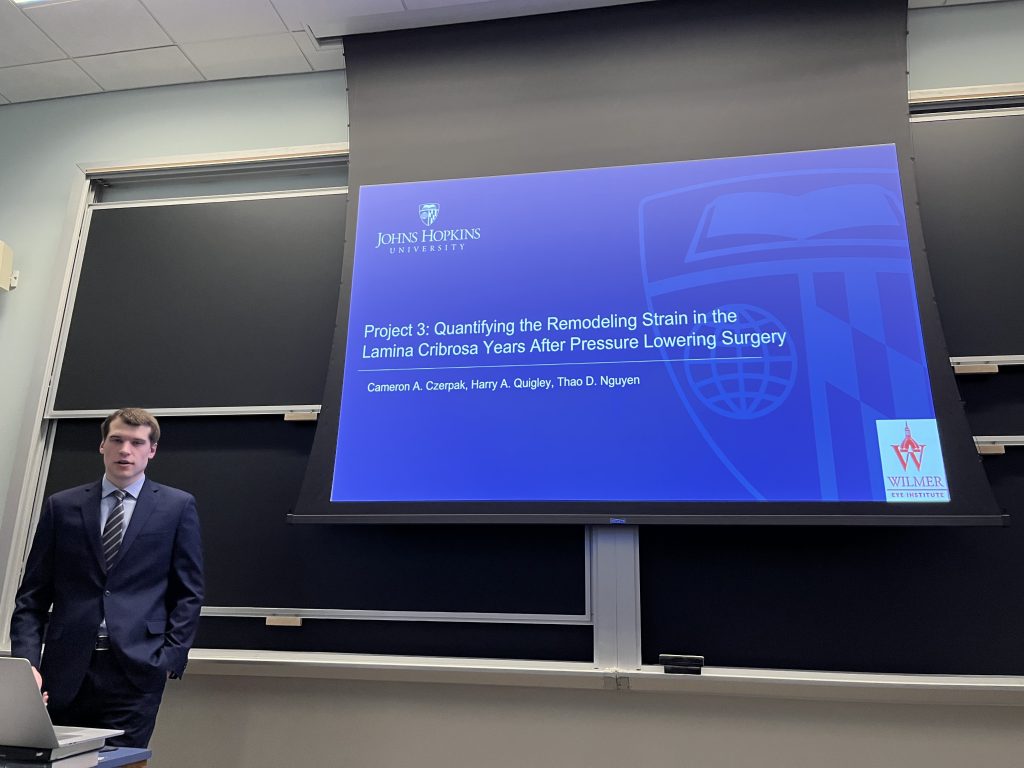
Cameron did a great job. His thesis came together beautifully.
Characterizing the Remodeling of the Lamina Cribrosa of Glaucoma Eyes
Glaucoma is the leading cause of irreversible vision loss and affects roughly 80 million people worldwide.
Glaucoma is characterized by progressive visual field loss from the death of retinal ganglion cell axons in
the optic nerve head (ONH). The primary site of axonal dysfunction in glaucoma is the lamina cribrosa
(LC), the part of the eye wall in the ONH. The human LC is formed from cribriform plates composed of
elastin, collagen types I and III, and various proteoglycans. When viewed en face, the plates resemble a
beam and pore network that supports axons exiting the eye. A major risk factor of glaucoma is excessive
intraocular pressure (IOP). High IOP mechanically deforms the LC, and those deformations stretch,
compress, and shear axons. Over time, the LC remodels and the plate-like structure thins and becomes
more curved with progressive axonal damage. The collagen, elastin, and proteoglycan composition also
change, and the pores become smaller. These changes in microstructure and tissue composition may
influence the biomechanics of the LC. LC biomechanics have previously been measured using digital
volume correlation (DVC) both ex vivo and in vivo for short term IOP change. However, the LC
deformations from long term remodeling have not been studied. The strain and remodeling may be
biomarkers for a patient’s susceptibility to glaucoma damage or worsening of vision loss following medical
intervention. Therefore, the objective of this work was to characterize the long-term remodeling of
the lamina cribrosa of glaucoma eyes by measuring LC biomechanics and structure.
The first part of this work characterized remodeling of the LC of postmortem human donor eyes and
compare to the degree of axonal damage. The second study estimates the direct strain response to IOP
change in vivo in glaucoma patients and their relationship to clinical measurements of glaucoma damage.
The final study reimages the glaucoma subjects of the second study years later to estimate the remodeling
deformations in the LC following medical intervention.


