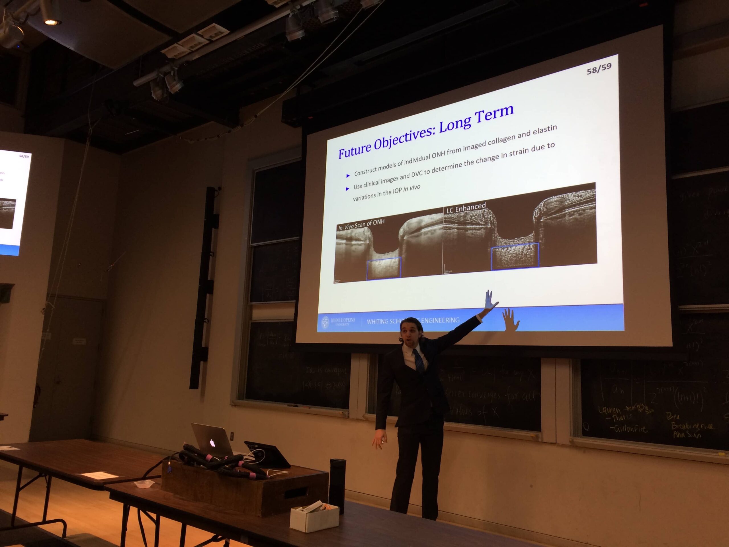
Congratulations to Dr. Dan Midgett for his successful dissertation defense, “Measuring the Full-Field Inflation Response of the Optic Nerve Head“. Abstract below.
The optic nerve head (ONH) is the region where the retinal ganglion cell (RGC) axons travel through the eye wall and bundle to form the the optic nerve. The lamina cribrosa (LC) and the peripapillary sclera (PPS) are the primary load-bearing structures of the ONH, supporting the RGCs and maintaining the pressure gradient between the intraocular and intracranial pressures. The LC is comprised of a network of collagen beams that wrap around the RGC bundles as they insert through the eye wall. The PPS is the stiff, thick portion of the posterior sclera that surrounds the LC. In the disorder known as glaucoma significant remodeling occurs in the tissues of the ONH, with changes in the collagen, elastin, and glycosaminoglycan (GAG) microstructure. This is associated with visual field loss and the death of RGC axons. While the mechanisms leading to vision loss remain unknown, glaucoma is commonly associated with older age and the level of intraocular pressure (IOP). A current hypothesis asserts that damage to the RGC axons is incurred mechanically through IOP-induced strains within the LC. If this hypothesis is correct, then the biomechanics of the LC and PPS are major contributing factors to the susceptibility of individuals to glaucoma damage. The objective of this work is to measure the three-dimensional (3D), pressure-induced deformation of the LC and PPS, and to investigate how the pressure-induced strains within the LC and PPS vary with region, age, LC shape, and GAG content.
We developed an ex vivo inflation test for the human posterior sclera, designed to measure the deformation of the ONH due to applied changes in the pressure within 21 eyes with no history of glaucoma. The pressure was varied between 5 and 45 mmHg, and images of the collagen and elastin within the LC and PPS were acquired using second harmonic generation (SHG) and two-photon fluorescence (TPF). Digital volume correlation (DVC) was used to calculate the deformation of the elastin and collagen between pressures, and analytical methods were developed to calculate the 3D strain fields within the LC and PPS at the resolution of the collagen beams within the LC (< 1μm). This study revealed heterogeneous strain fields with many localized regions of high strain. We found that the LC strains were smaller in older eyes and in the nasal quadrant, and larger on the periphery of the LC. We showed that this pattern of strain was consistent with the patterns of early glaucoma damage. Interactions between the LC and the PPS were also observed, with many eyes experiencing regions of compressive radial strain in the PPS adjacent to the LC. We also observed a significant reduction in the strains of the LC after the degradation of the majority of sGAGs within the LC and sclera. This demonstrates that the presence of sGAGs has a significant effect on the on the biomechanics of the ONH. This method was similarly applied to measure the IOP-induced deformation of the ONH within mouse eyes. Strains within the mouse LC were primarily nasal-temporal in magnitude and were significantly reduced after 1-3 days of chronic IOP elevation. LC strains were also smaller in older mice. We conclude that there are significant regional and individual variations in the susceptibility of the LC and PPS to strain induced through pressure. These variations may contribute to individual susceptibility to glaucoma.

