 Ocular injuries from blasts have increased in recent engagements caused by increasing prevalence of improvised explosive devices (IEDs). Such injuries are classified as primary and secondary injuries and are caused respectively by blast over pressures and fragments from debris. Several factors are hypothesized to influence the severity of primary eye injury in the literature: the high pressure shock front and the subsequent wave of lower sub-atmospheric pressure, threshold overpressure, and reflection of the shock wave by the orbit. There is, however, a dearth of clinical data that could verify the hypotheses above and establish the mechanism of the injury. Measuring and assessing the influence of these factors is difficult because survivable primary blast injuries are likely accompanied by injuries from fragments and blunt-force trauma and are thus more difficult to distinguish and enumerate. Moreover, the severity of the blast injuries and distance of the tertiary care facility from the injury site means that often patients are unable to recount the injury event, and witnesses are unavailable. The same limitations hinder clinical studies of the effectiveness of current eye armor, developed for ballistic and laser protection, in preventing blast injuries.
The overall goal of this project is to develop an experimentally validated computational model of the eye and apply the model to evaluate the stresses and deformations incurred by the eye-wall and critical ocular components from blast overpressures, and to investigate the interaction between the standard issue eye armor and the blast wave, and its effect on the mechanical loading of the eye. The model will be developed based on the following working hypotheses. 1) The anisotropic mechanical properties of the cornea and sclera as derived from the collagen structure, are critical to modeling the interaction of the blast wave and the globe. 2) The mechanical behavior of the cornea and sclera under dynamic (high-rate) loading is significantly different than the under quasistatic (slow-rate) loading. 3) The surrounding environment of the globe, including the extraocular tissues of the orbit and bony facial features is important in determining the effects of blast loading on the eye.
In addition to computational models, we are developing dynamic inflation experiments to characterize the high-rate behavior of the eye-wall and tissue simulates. The experiments uses 3D-DIC and a high-speed camera setup to capture the full-field elastodynamic displacement and strain response, then apply both analytical and computational models of structural vibration to determine material properties.
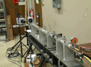
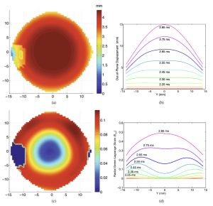
Figure 1 Dynamic inflation test using a shock tube, high speed imaging, and 3D digital image correlation (DIC). (a) Contours for the out-of plane displacement at t=2.45 ms and (b) line plots of the out-of-plane displacement for each 0.1 ms interval from 2.05 – 2.85 ms. (c) Green- Lagrange strain at t = 2.45 ms for the same specimen and (d) radial strain at X = 0 for each 0.1 ms interval from 2.05 – 2.85 ms.
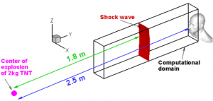
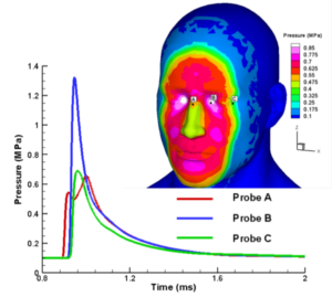
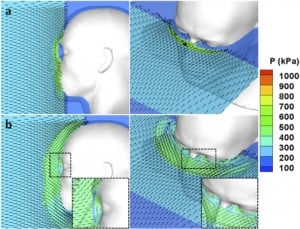
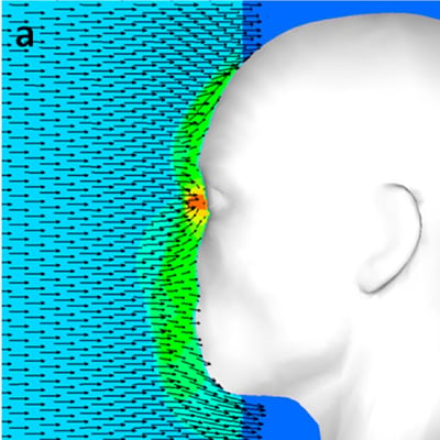
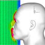
Figure 2: Computational modeling of blast impact on a rigid face comparing the effects of wearing googles and spectacles on blast overpressure. Without eye protection, the blast overpressure for a blast directly in front of the face is highest at the eyes.
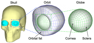
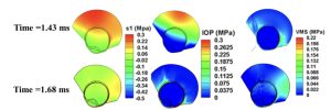
Figure 3: (a) Computational model of blast propagation through a deformable eye in a rigid skull. (b) The model for the eye is surrounded by orbital tissues and contains the major intraocular tissues, such as the lens, retina, and optic nerve head. (c) Contours of the maximum principal stress, intraocular pressure, and von Mises stress at two different time points showing wave reflections produces high tensile stresses in then posterior eye wall and eye rotation causes large von Mises stress at the attachment of the eye wall with the orbital tissues.
Collaborators
- Rajneesh Bhardwaj, PhD – Assistant Professor, Department of Mechanical Engineering, IIT Bombay
- Kaliat T. Ramesh, PhD – Alonzo G. Decker Jr. Professor of Science & Engineering, Department of Mechanical Engineering, Johns Hopkins University
- Oliver Schein, MD – Burton E. Grossman Professor of Ophthalmology, Director, Comprehensive Eye Service, Cornea and Anterior Segment Service, Johns Hopkins Medical School
- Gehard Grimm, Adam Fournier PhD – Army Aberdeen Test Center.